Swellings of the salivary glands can be signs of
inflammation, obstruction by stones or strictures, tumors and other causes

Anatomy of the salivary glands of the head:
parotid gland (pg), submandibular gland (gsm), sublingual gland (gsl)

Computertomography image of a calcified tumor
of the left submandibular gland (arrow)

Introducing an endoscope for salivary ducts (sialendoscope)
with the help of a so-called port. Simple diagnostic measures can be even
carried out without any anesthesia.

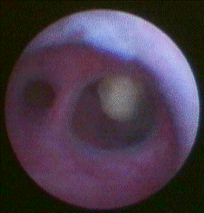
Endoscopic view of a stone in the duct system
of the parotid gland (Stensen's duct) behind a branching.

A basket is introduced through the working
channel of the endoscope to grasp and remove the stone.

Tip of a sialendoscope with an inlying basket
that has captured a stone.

Tips of sialendoscopes with a miniforceps (top),
laser fiber (middle) and drill (bottom) for the fragmentation of salivary
gland stones.

Endoskocopic view of a laser light impulse
being applied to a salivary gland stone (laser lithotripsy)

Endoscopic view of a salivary gland stone
which was fragmented by laser lithotripsy.

Fragments of stones which were broken and
removed by forceps under sonographic control (sonoguide forceps)

Fragmentation of salivary gland stones
through acoustic shock waves (extracorporeal shock wave lithotripsy). This
method is usually applied in local anesthesia without the need of any
cutting.

Small stone fragments which left the salivary
ducts after extracorporeal shock wave lithotripsy.

Endoscopic view of a stricture of a salivary
duct.

Also balloons can be sled through the working
channels of sialendoscopes. They can be used to dilate strictures.

A drain is mounted on a sialendoscope so that
it can be placed in the duct system under endoscopic control to prevent the
development of new strictures after dilatation.

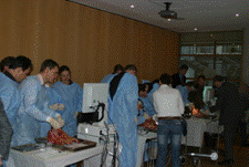
In case that you should be a physician
interested in the techniques described here: We have organized and taught on
several practical and theoretical instruction
course during national and international conferences. If you should be
interested in these please either contact us or search on the websites of
scientific medical associations.

Welcome...
...to
our site on the diagnosis and treatment of diseases of the salivary
glands with special focus on obstructive diseases (salivary gland stones
and strictures). We hope that this sites provides interesting
information for you and we would be happy to receive suggestions for
improvements or other feedback.
Urban Geisthoff
M.D. / Prof. Dr. med.
for the
salivary gland treatment centre in
the Department for Otorhinolaryngology / ENT, Head and Neck Surgery of
the University Hospital of Marburg (Chairman: Prof. Dr. B.A. Stuck) , Germany
Please note...
The information on this site can not
replace medical advice or treatment (see also disclaimer).
We restricted the topics on this site for reasons of clarity:
There exist also other techniques than those described here.
Additionally, there is a multitude of other conditions, disorders and
diseases which can affect the salivary glands (e.g. Sjögrens syndrome or
recurrent juvenile parotitis) which is not covered by this site for the
same reason.
What are the typical signs of stones and strictures (obstructive disease),
and of tumors?
The
typical signs of obstructive disease are swellings during meal-time. The
regions mainly affected are the cheek (parotid gland) or the area below
the chin (submandibular gland). Additional pain can be a sign of
infection (sialadenitis). Tumors (benign and malign neoplasms) are often
associated with chronic swellings which are frequently painless. The
nerve for the movement of the face has a close contact to the salivary
glands. If the motion of the face should be affected you should visit a
physician soon.
What
examinations can be done for diagnosis?
Clinical examination and a ultrasound
examination of the head and neck with a high-end machine are often
sufficient for a diagnosis to diagnose tumors, stones and inflammatory
disease. Sometimes it might be useful to perform an endoscopy of the
salivary duct system (sialendoscopy, sialoscopy). Further methods are
magnetic resonance imaging (mri) or sialography, computer tomography (ct
scan) and very seldom szintigraphy. In case that the findings are
suspicious of a tumor it can be helpful to perform punctions for fine
needle aspiration cytology or even core bore biopsies.
What treatment options exist?
Tumors
are typically treated by total or partial removal of the gland from the
outside. Main priorities after total removal of the tumor are the
preservation of the facial nerve and aesthetic aspects. Magnifying optic
systems like loops or microscopes and electrophysiologic monitoring
methods are often used to avoid damage to the nerve.
In case of small stones it might be worthwile to stimulate saliva
production and wait for a while as some stones will leave the duct
system spontaneously. Otherwise a multitude of different
minimal-invasive options has emerged in the last two decades and this
development seems to continue.
In
former times the gland had to be remove in all cases. An alternative is
the surgical removal through the mouth in many cases. This is sometimes
possible with classical surgical instruments. In a number of cases
it is now also possible to reach the stones with small endoscopes
through the natural orifices of the ducts. These so-called
sialendoscopes have outer diameters of about 0.6 to 2.0 mm. They have
working channels to insert instruments for stone retrievel like baskets,
graspers and miniforceps. If the stone should be to large an attempt of
fragmentation with instruments like lasers or drills can lead to success.
Instead of using endoscopic control it is sometimes also possible to use
forceps guided by ultrasound for fragmentation.
A further option is the use of acoustic
shock waves. This technique is called extracorporeal shock wave
lithotripsy (ESWL). The focus of the shock wave is centered into the
stones. Small fragments can be washed out by the natural flow of saliva.
Sometimes several sessions are necessary. The combination of different
techniques is often useful and increases the chances of success (so-called
multimodal therapy).
Strictures can also be treated under
endoscopic or ultrasonographic control with instruments like balloons,
drills, forceps and drains. Like for stones the combination of different
techniques is often useful.
Contact
For
further questions or if you are interested in an appointment at our
out-patient clinics for salivary diseases please address:
Secretaries
Dr. Urban Geisthoff
M.D./ Prof. Dr. med.
(further information regarding his qualifications is published under
www.geisthoff.de)
Salivary gland centre, ORL/ENT department,
(Chairman: Prof. Dr. B.A. Stuck)
University Hospital Marburg
Postal adress:
Prof. U. Geisthoff
Univ.-HNO-Klinik
Baldingerstr.
35043 Marburg
Germany
via
in Englisch:
Ms. Susanne Zapf (lead of the International Office)
Tel.: + 49 (0) 6421 58 66808
Fax: + 49 (0) 6421 58
96111
Email:
susanne.zapf@ukgm.de
in German:
Secretaries (Ms. Hinkelmann, Ms. Luppolo, Ms. Bender, Ms. Oetzel)
Tel.: + 49 (0) 6421 58 66603
Fax: + 49 (0) 6421 58 66367
Email:
sekretariat.hno.mr@uk-gm.de
Please
note that the regulatories for German physicians limit the amount of
information by telecommunication nets (including telephone, email,
internet etc.).
Further information on the topic and on us can also be found under the position "Travel
/ links" .
What is a salivary gland centre?
The
term "salivary gland centre" is neither protected nor well defined.
Therefore this is an additional information about a specialization
according to our own judgement. We
think that it is probably adequate to use it for our facility as we
- have
broad
experience on this topic for a long time now.
- can
offer the whole spectrum of different diagnostic an therapeutic options
in cooperation with the other departments of our hospital.
- are
equipped with all instruments necessary for treatment. These sets of
instruments were chosen from different suppliers so that we can cover
various different situation in an optimal way.
-
are doing research in the field. We contributed to the development of
special instruments and new techniques. We were able to present these on
conferences and publish these in the literature.
-
instructed other physicians on this topic during medical courses during
scientific conference in Germany and abroad.
- are treating not only patients from
the region but also from other countries.
- take
part in the development of medical guidelines.
-
offer a special outpatient clinics for salivary gland diseases.
- have
just a passion here.
Addendum
Of course there are a lot of other
qualified colleagues. To find these you can ask your general
practitioner, other physicians treating you, the chambers of physicians
("Aerztekammern"), scientific medical associations.
We
would like to list a selection of some of those which we know. Please do
understand that this list for sure not complete, and that our recommendation can never be a guarantee but is for
information only and without responsibility.
-
Prof. Dr. H. Iro and coworkers in Erlangen, Germany
-
Prof. Dr. O. Guntinas-Lichius and coworkers in Jena, Germany
- Prof. Dr. J. Zenk and coworkerst in
Augsburg, Germany
- Dr.
Ph. Katz and coworkers in Paris, France
-
Prof. Dr. M. McGurk and coworkers in London, UK
- Dr.
F. Marchal and coworkers in Geneve, Switzerland
-
Prof. Dr. O. Nahlieli and coworkers in Ashkelon, Israel
-
Prof. Dr. M. Fritsch and coworkers in Indianapolis, USA
Of course you are also invited to visit
our
-
salivary gland treatment centre in Marburg (please
find contact data of Dr. Geisthoff above).
We do not take responsibility for the
content of linked external web sites.